TAVI (transcatheter aortic valve implantation)
Replacing a narrowed aortic valve through an artery in the leg.
What it is
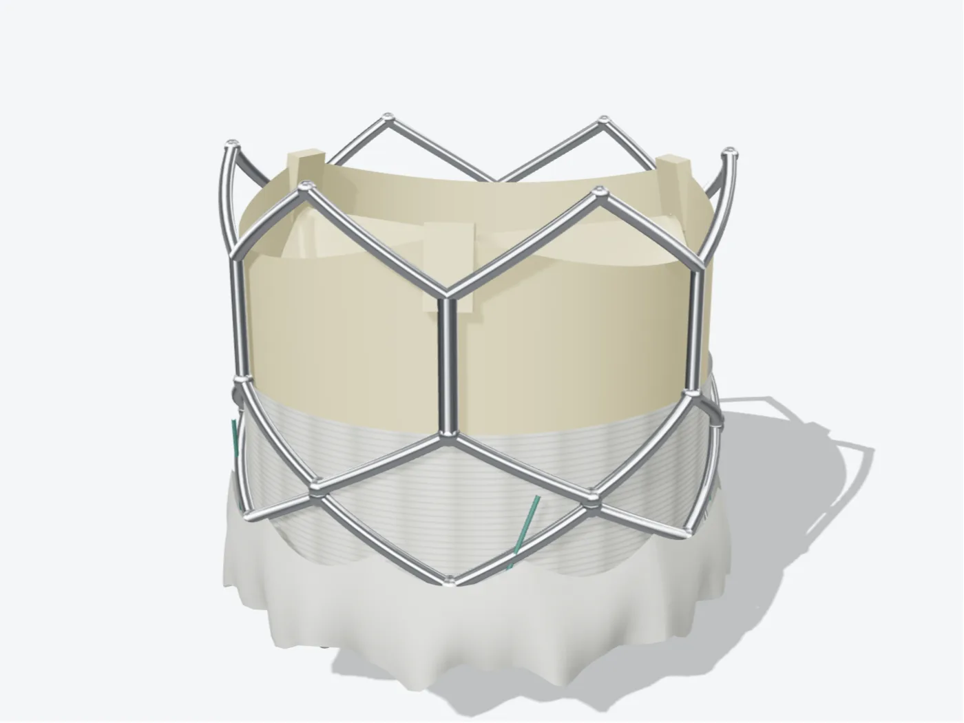
TAVI places a new aortic valve inside the old, narrowed one. The new valve is folded onto a catheter, passed up from an artery in the groin and expanded inside the diseased valve, where it starts working straight away. It avoids open-heart surgery.
Why it may be recommended
- Severe aortic stenosis causing symptoms
- A heart team recommendation that TAVI suits you better than surgery
Planning · CT
The route to the heart, measured before the day.
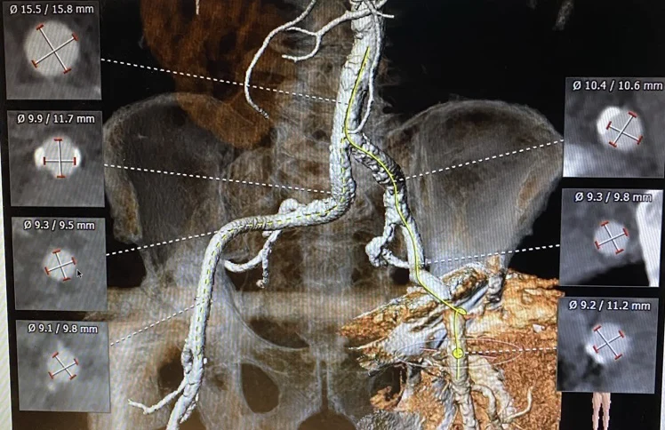
Before TAVI, a CT scan maps the arteries from the groin up to the heart. The planned route is traced along the artery and its width is measured at each point, so the team can check the valve can travel that way and choose its size.
- Checkpoints
- 6Cross-sections along the route
- Widest point
- Ø 15.8 mmMeasured across two axes
- Narrowest point
- Ø 9.1 mmThe tightest part of the route
How TAVI works, step by step
Step 1 of 4
A CT scan measures the valve and the arteries so the right valve size is chosen.
Under sedation and local anaesthetic, a catheter is placed in the artery at the groin.
The folded valve travels up to the heart and is positioned inside the narrowed valve.
The new valve is expanded and pushes the old leaflets aside. It works immediately.
Before
- Echocardiogram, CT scan and a coronary angiogram
- A dental check, as valve infections often start in the mouth
- A discussion with the heart team
Recovery
- Usually one to three nights in hospital
- Walking the next day for most people
- Echocardiogram follow-up of the new valve
Risks
TAVI carries risks including bleeding or damage to the artery, stroke, the need for a permanent pacemaker, leakage around the valve and, rarely, more serious complications. These are weighed carefully against the risks of not treating severe aortic stenosis.
Considering this procedure?
Every procedure is discussed with you first: why it is recommended, the alternatives, and the risks that apply to you.