Left atrial appendage occlusion
A small plug that lowers stroke risk in atrial fibrillation without lifelong blood thinners.
What it is
In atrial fibrillation, most stroke-causing clots form in a small pouch called the left atrial appendage. An occlusion device is placed in the opening of this pouch through a vein in the leg, sealing it off. It is an option for people who cannot safely stay on long-term blood thinners.
Why it may be recommended
- Atrial fibrillation with a high stroke risk
- Bleeding problems, or a high bleeding risk, on blood thinners
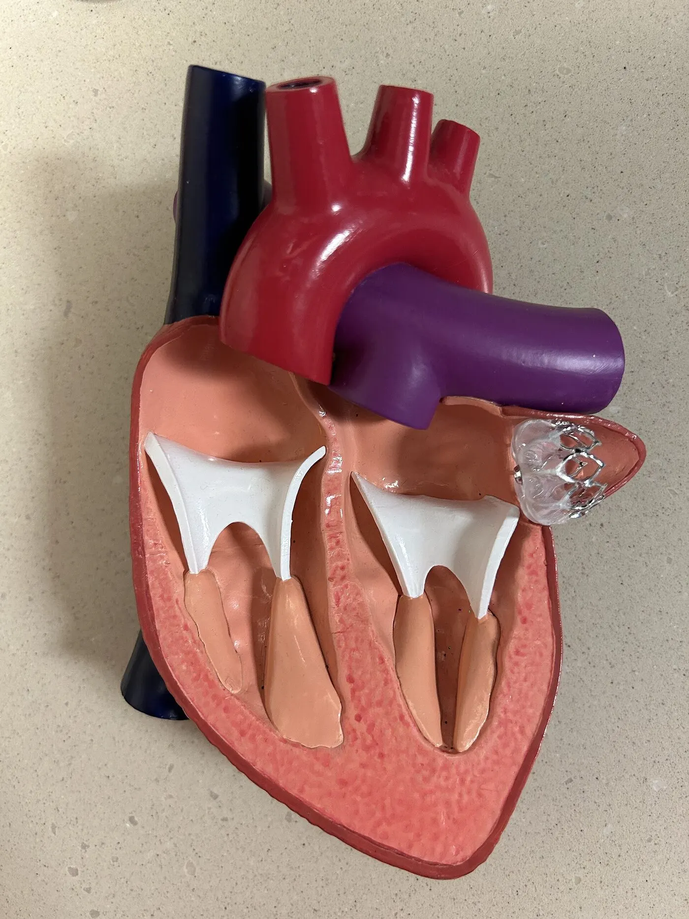
How laa occlusion works, step by step
Step 1 of 4
A CT scan or transoesophageal echo measures the appendage.
Under anaesthetic, a catheter passes from the leg vein to the left upper chamber.
The device opens in the mouth of the appendage like a small umbrella.
Its position and seal are checked with ultrasound before release.
Before
- CT or transoesophageal echo
- A plan for your blood-thinning medicines
- Fasting beforehand
Recovery
- Usually one night in hospital
- Short-term blood-thinning tablets
- A follow-up scan to confirm the seal
Risks
Risks include bleeding, fluid around the heart, device movement, clot on the device and, rarely, stroke. Dr Ramzy will discuss how these compare with your risks on medicines alone.
Considering this procedure?
Every procedure is discussed with you first: why it is recommended, the alternatives, and the risks that apply to you.