Coronary angiogram
X-ray pictures of the heart arteries, taken through a small tube in the wrist.
What it is
A coronary angiogram uses a thin, flexible tube (catheter) and X-ray dye to show the inside of the coronary arteries. It is the most accurate way to find out whether an artery is narrowed, and if so, how much.
Why it may be recommended
- Angina or chest pain thought to come from the heart
- An abnormal CT coronary angiogram or stress test
- Before valve treatment such as TAVI
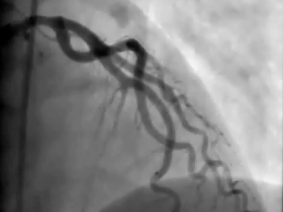
How angiogram works, step by step
Step 1 of 4
You are awake but comfortable. The skin over the artery at the wrist (or occasionally the groin) is numbed.
A thin tube is guided up to the heart. You do not feel it moving inside the artery.
A small amount of X-ray dye shows each artery on the screen. You may feel a brief warm flush.
If a narrowing is borderline, a pressure wire or imaging catheter measures its true effect before any decision.
Before
- Fasting for a few hours beforehand
- A list of your medicines; some blood thinners or diabetes tablets may be paused
- Someone to drive you home
Recovery
- A firm band on the wrist for a few hours
- Most people go home the same day
- Avoid heavy lifting with that arm for a few days
Risks
Serious complications are uncommon. Possible risks include bruising or bleeding at the puncture site, reaction to the dye, kidney strain and, rarely, damage to an artery, heart attack or stroke. Dr Ramzy will talk through what applies to you.
Considering this procedure?
Every procedure is discussed with you first: why it is recommended, the alternatives, and the risks that apply to you.